Radiologic technologists – typically known as radiographers – perform diagnostic medical imaging examinations. The images that radiographers capture are utilized by physicians so that they may create proper treatment plans for their patients. They can be employed in a number of settings, including hospitals, free-standing outpatient imaging centers, and even prisons. What may not be as commonly known is that radiographers can specialize in several forms of the practice.
We asked the experts at Pima Medical Institute about some of the more popular choices for a radiographer. Here’s what they had to say:
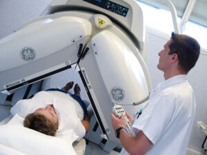
 1) Computed Tomography (CT)
 1) Computed Tomography (CT)
Computed tomography (CT) involves taking images, or “slices” of a patient’s anatomy using a rotating X-ray unit. These images allow radiographers and physicians to more closely view the internal anatomy of their patients. The CT images are produced and assembled slice by slice in order to create a full picture of the internal organs.
2) Magnetic Resonance Imaging (MRI)
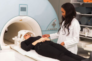 2) Magnetic Resonance Imaging (MRI)
During an MRI scan, the radiographer applies a radiofrequency pulse to a magnetic field surrounding the patient’s body. The atoms within the patient’s body are knocked out of alignment during the pulse. When it’s turned off, the atoms return to their original position. During the process, the atoms give off signals, which are measured by a computer and processed into detailed images of the patient’s anatomy.
3) Bone Densitometry
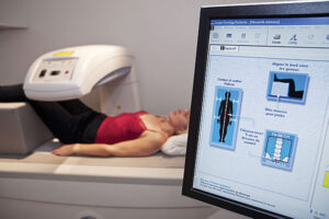 3) Bone Densitometry
In bone densitometry, the radiographer measures bone mineral density at certain areas of the patient’s body, such as the wrist, spine, heel or hip to calculate total body bone mineral content. The exam can measure the rate of bone mineral loss over a specific period of time, which helps estimate the patient’s potential risk for fractures.
4) Mammography
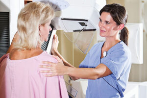 4) Mammography
Mammography is used to detect the onset of breast cancer. Mammographers produce diagnostic images of breast tissue using X-ray equipment, which provides excellent detail and information. They must be accurate with their placement of the equipment and exposure techniques in order to obtain a clear image while delivering as low of a radiation dose as possible to maintain high safety standards.
 5) Nuclear Medicine
 5) Nuclear Medicine
Nuclear medicine technologists utilize radioisotopes (radioactive substances placed inside the body) to obtain information about organs, tissues and bones. Once the substance is given to the patient, the nuclear medicine technologist uses a special camera to detect gamma rays that are emitted by the radioisotopes to create an image of the body part being examined.
Pima Medical Institute offers the Radiography program at the following campuses: Mesa and Tucson, Arizona, Chula Vista, California, Denver, Las Vegas, Albuquerque, New Mexico, Houston, and Seattle. Classes focus on a number of topics, including the principles of exposure, radiographic biology, pathology and more. Once students have completed the program, they may sit for the American Registry of Radiologic Technologists’ Radiography Certification Exam.